EARLY MICROSCOPES IN SOUTH AUSTRALIA
Website for the Virtual Museum
Home
Coming meetings
Past meetings
About the Society
Main Galleries
Medicine
Surgery
Anaesthesia
X-rays
Hospitals,other organisations
Individuals of note
Small Galleries
Ethnic medicine
- Aboriginal
- Chinese
- Mediterran
EARLY MICROSCOPES IN SOUTH AUSTRALIA
Acknowledgements: We are most grateful to Dr. Peter Hetzel, the President of the Royal Adelaide Hospital Heritage Society, and Margo Way, the Manager of their Museum. They allowed us to photograph some of the early microscopes. We also thank the management of the South Australian Royal Society for permission to photograph some of their microscopes. Dr. P. W. Allan allowed us to photograph recent microscopes at the Flinders Hospital. John Brearly kindly allowed us to photograph the electron microscope at the IMVS in Adelaide. We are also most grateful to Professor Donald Simpson for allowing us to view Hooke's Micrographia and for his valuable suggestions.
Additional information about Lister's father, Verco, Ernst Ruska and other aspects of light and electron microscopy are mentioned in the appendix.
Background: Before the advent of the microscope in the 17th century, the periodic incidences of epidemics were attributed to "miasma" (defined in the OED as "infectious or noxious exhalations"). The most dangerous times were in the summer, and the more affluent members of the population tried to avoid illnesses by leaving the cities during the warmer periods, and moving into the country or higher, cooler regions.
The invention of the microscope enabled scientists to see bacteria and establish a connection between their presence and illness. The earliest use of magnification is mentioned by Seneca and Pliny. It seems that transparent crystals, particularly if convex in shape, could magnify or focus sun rays to produce heat. "Reading stone" or "fire stone" was the names sometime given to such crystals. The development of eyeglasses (spectacles) followed, and by 1500 AD these were quite common as lens technology developed. The word lens appears to be derived from the Latin word "lens", meaning lentil, due to the similar shapes of a biconvex lens and a lentil seed.
In 1590, the Dutch spectacle makers Zaccharias Janssen and his son Hans, experimented with several lenses in a metal tube, and achieved considerable magnification.
Anton Van Leeuwenhoek (1632-1723) is considered as the father of microscopy because he was the first to notice moving organisms, red cells, and bacteria. His later instruments (1673) magnified 200 fold.
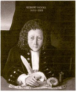
The English contribution was provided by Robert Hooke, who was a Secretary of the Royal Society. He published in 1665 a paper named Micrographia subtitled “Minute Bodies” Using a 30 fold magnification he described rectangular structure in a piece of cork and may have been the first to use the word “cell”. Further improvements followed.
Chromatic aberration was recognized and corrected, magnification was increased to 1000 fold, and several magnifying lenses could be attached to a single tube by a rotating galley. Binocular microscopes, and projection and photographic options are fairly recent developments.
Australia did not became an efficient scientific society until the mid 19th century. However, communications with Europe and the USA were quite efficient. The first X-rays appeared in Australia within a year of Röntgen's discovery in 1896. It is therefore certain that microscopes were available before then.
Sir Joseph Verco (please see appendix) became a lecturer in Medicine and Therapeutics at the Adelaide University in 1887, when the Clinical School opened. He brought with him from England several microscopes. These are now stored at the Adelaide Royal Society and in the RAH Museum.
The following photographs represent some of these early microscopes and others used today.
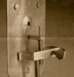
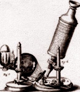
TWO EARLY MICROSCOPES, c. 1670. ON THE LEFT IS THE FIRST VAN LEEUWENHOEK MODEL. IT HAS ONLY ONE LENS. SCREWS ARE USED FOR FIXATION OF THE SPECIMEN, WHICH IS ATTACHED TO THE PIN. A SIMILAR IVORY MODEL IS ON THE RIGHT.
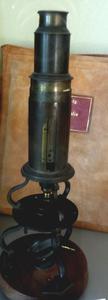
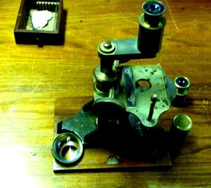
AN EARLY MICROSCOPE BROUGHT TO ADELAIDE BY SIR JOSEPH VERCO IN 1878. THE SLIDES ON WHICH THE TISSUE SPECIMENS WERE PLACED ARE AT THE TOP LEFT CORNER, POSSIBLY MADE IN ENGLAND BY GEORGE DIXON, CIRCA 1870.
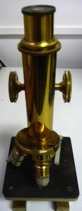
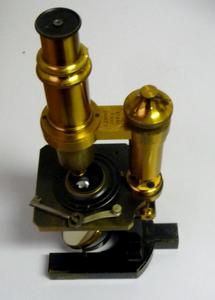
TWO MICROSCOPES ATTRIBUTED TO SIR JOSEPH VERCO.
ON THE LEFT IS A LATE 19TH CENTURY ENGLISH MODEL, STORED AT THE ADELAIDE ROYAL SOCIETY (RS). THE ONE ONE THE RIGHT IS A COMPOUND BRASS AND STEEL MODEL MADE BY CARL ZEISS IN JENA, CIRCA 1905. IT IS STORED AT THE RAH.
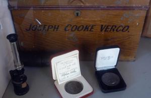
A SMALLER MICROSCOPE AND A BOX FOR SIR VERCO'S MICROSCOPES AND COMMEMORATIVE MEDALS, EXHIBITED IN THE FOYER OF THE RS. DR. VERCO MAY HAVE USED THE MICROSCOPE TO EXAMINE HIS CONCHOLOGY COLLECTION.
A LATER MICROSCOPE WITH THREE ROTATING LENSES (LEFT). THE REVOLVING NOSE PIECE IS SAID TO BE INVENTED BY ERNST LEITZ IN WETZLAR, GERMANY. THE MODEL HAS BEEN DATED TO 1905.
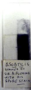
A SLIDE OF BACILLUS SUBTILIS BY SIR ALEXANDER FLEMING (RIGHT). IT IS POSSIBLE THAT IT WAS PROVIDED BY PROFESSOR ROWLEY. THE GLASS SLIDE AND A THIN COVERSLIP IS STILL USED TO PREPARE SPECIMENS FOR MICROSCOPY.
THE TISSUE SECTIONS ARE USUALLY 2-5 MICRONS THICK AND ARE CUT BY A MICROTOME.
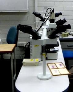
TEACHING MICROSCOPE AT THE FLINDERS MEDICAL CENTRE. PLEASE NOTE THE ADDITIONAL EYE PIECES AND CAMERA ATTACHMENTS.
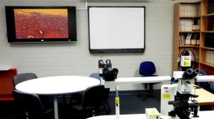
A MEETING ROOM IN THE SAME HOSPITAL. THE MICROSCOPE SLIDE CAN BE PROJECTED ON A TELEVISION SCREEN FOR THE AUDIENCE TO SEE. THE TISSUE SHOWN (TOP LEFT) IT IS UTERINE MUSCLE.
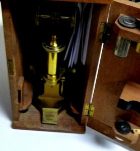
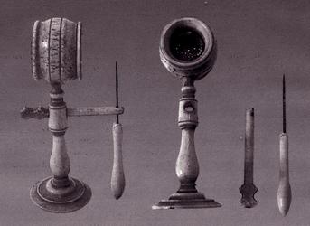
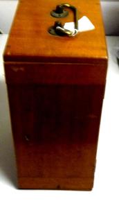
EARLY MICROSCOPES WERE PORTABLE AND KEPT IN WOODEN BOXES. OPEN AND CLOSED BOX IS SEEN ABOVE. AN EYEPIECE IS VISIBLE AT THE TOP OF AN OPENED DOOR.
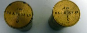
THE EYE PIECES WERE STORED IN METAL CONTAINERS MANUFACTURED IN LONDON BY THE BECK BROTHERS, NEPHEWS OF JOSEPH JACKSON LISTER, AN OPTICIAN AND WINE MERCHANT WHO WAS RESPONSIBLE FOR PRODUCING THE FIRST ACHROMATIC LENSES. JOSEPH JACKSON LISTER WAS THE FATHER OF THE NOTED SURGEON JOSEPH LISTER WHO INTRODUCED ASEPSIS IN SURGERY.
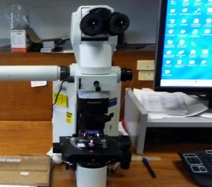
A CONTEMPORARY MICROSCOPE ON THE DESK OF A PATHOLOGIST, IT IS BINOCULAR, AND HAS A CAMERA FACILITY. THE COMPUTER SCREEN IS ON THE RIGHT, AND AN ADDITIONAL VIEWING ARM POINTS TO LEFT.
ELECTRON MICROSCOPES
The resolution of a light microscope is limited by the wavelength of light, and any feature smaller than 200 nanometres will be difficult to visualise accurately. The wavelength of a beam of electrons is much less, and its resolution is approximately 10 nanometres (1 nanometre = 0.001 microns = 0.000001 millimetres).
The first concept of an electron microscope was suggested by Ernest August Friedrich Ruska. He built an "electron lens" in 1931, and was awarded the Nobel prize in 1986. Ernst August Ruska was born in December 1906, the 5th of seven children. His father was a Professor of Asian Studies in Heidelberg. His early studies were at the Technical University in Munich, but in 1928 he moved to Berlin to work with Max Knoll. Even in high school he was aware that the resolution of light microscopy was limited by the wavelength of light, as calculated by Ernst Abbe (1865). He was also aware that the wave length of electrons was much less. Working with Max Knoll he produced the first "Electron Lens", which was an electromagnetic ring. It could focus electrons similar to an optical lens, but with a tenfold magnification, and rotated the electron beam around its axis. The amount the electron beam rotates increases as the magnification does.
In 1937 he joined the Siemens company in order to accelerate the commercial development of his invention. Later developments included the scanning, and tunnelling electron microscopes. Their contribution to molecular biology and physics were invaluable.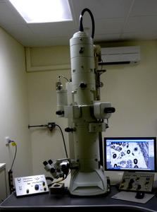
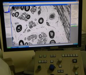
CURRENT ELECTRON MICROSCOPE USED AT THE ROYAL ADELAIDE HOSPITAL. DIGITISED DISPLAY OF A NERVE FIBRE IS ON THE RIGHT. THE OSMIUM STAINS THE MYELIN SHEATHS BLACK.
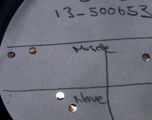
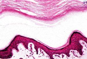
NERVE AND MUSCLE PREPARATIONS FOR THE ELECTRON MICROSCOPE SHEATHED IN COPPER, SEE LEFT ABOVE. THE SECTIONS ARE CUT WITH A DIAMOND KNIFE AND ARE ABOUT 200 NANOMETRES THICK. PHOTOGRAPH OF SKIN HISTOLOGY ON THE RIGHT.
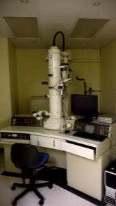
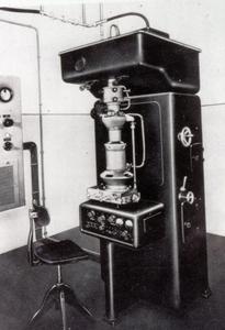
TWO OLDER ELECTRON MICROSCOPES: ONE ON THE LEFT WAS USED AT THE QEH IN THE 1970'S; THE ONE ON THE RIGHT WAS USED SOME 20 YEARS EARLIER. THE DIGITAL DISPLAY WAS NOT AVAILABLE THEN.
The tubular structure of the microscopes is necessary to accelerate the electrons toward the specimens at the bottom. The electrons are generated from heating a Tungsten element at the top. An internal vacuum is needed to reduce the Tungsten wastage, and to prevent the electrons interfering with other particles. A potential difference of 10 - 200 kV then accelerates the electron toward the specimen.
The De Broglie wavelength of the electrons is inversely proportional to the accelerating voltage, as show below:
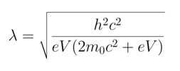
which can be approximated as:
Where:
De Broglie wavelength of the electrons Rest mass of an electron e Charge of an electron h Planck's constant c Speed of light V Accelerating voltage
APPENDIX:
WE THOUGHT THAT SOME OF THE NOTED PERSONS MENTIONED IN THE PRESENTATION NEEDED FURTHER COMMENTS.
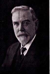
Sir Joseph Cook Verco was born in Adelaide in 1851. There was no medical school in South Australia, so, with others, he travelled to London to study medicine. He graduated MRCS in 1873 and gained numerous other degrees: MB London and LRCP 1876, FRCS and BS (Gold Medal) 1877. His clinical experience was gained at St. Bartholomew's Hospital. He returned to Adelaide and obtained a MD. He registered with the Medical Board in May 1878 (registration number 261). His clinical appointment was at the Royal Adelaide Hospital as a visiting Physician, but when the Medical school was established he became Lecturer in Medicine and Therapeutics. He served on numerous public, University and BMA Committees, and was a member and later President of the Royal Society of Adelaide. His main non medical interest was in Conchology. Between 1890 and 1911 he published 26 scientific papers on the topic.
His microscopes are a part of a larger collection gathered by Professor James McCartney, a Bacteriologist at the University of Adelaide in the 1950's. Histology and tissue diagnosis were an important part of the medical curriculum. Thus many Australian medical students, before the Adelaide medical school was established, bought their microscopes in England and brought then back to Australia after graduation.
Dr. John Britcher was involved with preserving these, and currently most of them are stored at the Royal Adelaide Historical Society museum.
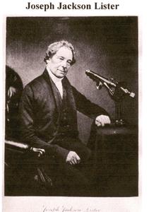
Joseph Jackson Lister was born in 1786 and was apprenticed to his fathers wine business. At the age of 18 he became a partner. J. J. Lister was interested in nature and in microscopy. He found the early microscopes had considerable chromatic defects, and he set out to produce corrective achromatic lenses. He published his work in 1830 in a paper entitled "On some properties in achromatic object-glasses applicable to the improvement of the microscope". He also commissioned William Tulley to produce an improved microscope and a stand. His activities resulted in a Fellowship of the Royal Society in 1832.
He had six children: Mary, John, Isabella Sophia, Joseph, William Henry, and Arthur Hugh.
Joseph, the third born, later became Lord Lister known for his contributions to antisepsis.
Robert Hooke, (1635-1703). There does not appear any authenticated painting of Hooke, the above is an artists impression. Hooke's interests were in physics and chemistry. His Alma mater was Christ Church, Oxford; and in his early years worked with Robert Boyle. He is known to have produced some vacuum pumps for Boyle's experiments. He is known for establishing the Hooke's law (F=kX) which involves the forces needed to change spring tension and is relevant to the performance of barometers, spring gauges and watches. He became the curator for experiments of the Royal Society in 1662 and later its secretary. Hooke's other interests involved microscopes and telescopes. His descriptions of the use of the microscope are summarised in his publication of "Micrographia" where he describes his findings using a microscope. His description of the cork may have coined the name "cell".
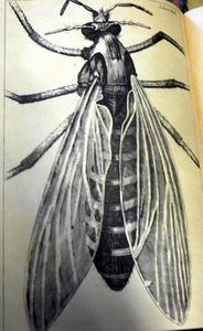
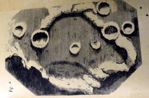
The above are the drawings of Hooke's findings. His microscope probably had a magnification of 30 or more. The light source for his microscope could have been a paraffin lamp. He also used a telescope, and his picture of the moon's craters is shown above.
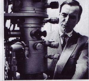
PHOTOGRAPH OF ERNST RUSKA WITH HIS MICROSCOPE
Ernst Ruska was born in 1906 in Heidelberg. His early education was at the Technical Universities in Munich (1925-1927) and later in Berlin where he obtained his PhD. His Doctoral adviser was Max Knoll. Even then he proposed that as the ordinary light waves limited the resolution of optical microscopes (Abbe's formula), and that electrons with a wave lengths perhaps even 1000 times shorter if "focused" could produce much greater magnifications. In 1931 he showed that a magnetic coil could act as an electron lens and he used several such coils to make a first electron microscope in 1933. The commercial application needed private enterprise and this was provided by Siemens, and the first such electron microscope was produced in 1939. The benefits to biological sciences and physics were incalculable. Ruska's brother Helmut, a medical doctor, was involved the medical applications.
Ruska left Siemens in 1955, and became a director of electron microscopy at the Fritz Haber Institute. He also taught at the Technical University of Berlin from 1957 until his retirement in 1974. He was awarded half of the Nobel prize in 1986. The other “quarter” prizes were given to Gerd Binning and Heinrich Rohrer for their design of the scanning/tunnelling microscope.


-o0o-